スポンサードサーチ
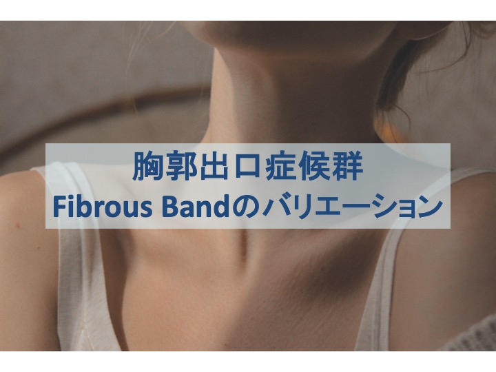
胸郭出口症候群
『胸郭出口症候群(Thoracic Outlet Syndrome:TOS)は、胸部上部領域で、鎖骨下動脈・静脈と腕神経叢への圧迫によって特徴づけられている病態です。
胸郭出口症候群の診断と治療は、非常に複雑となっています。そのため、まずは胸郭出口症候群の病態を引き起こす原因を知る必要があります。

胸郭上部において圧迫を生じさせる因子は、骨組織と軟部組織関連の異常が含まれます。
『骨組織の要因としては、頸肋、異常な第一肋骨、長い第7頸椎横突起などが含まれます。一方で軟部組織の要因としては、靭帯、線維帯(Fibrous Band)、斜角筋の異常が存在し、これらにより圧迫され症状を引き起こすと考えられます。
- 頸肋
- 異常な第一肋骨
- 長い第7頸椎横突起
- 靭帯
- 線維帯
- 斜角筋の異常
このような異常な解剖学的な構造は、腕神経叢の機械的圧迫・鋭い刺激など基本的な問題を引き起こす可能性があります。
胸郭上部領域は解剖学的バリエーションが豊富であるため、今回の記事では『胸郭上部におけるFibrous Bandのバリエーションと臨床上の注意点』をご紹介していきます。
Fibrous Bandのバリエーション
先ほどから何度か登場している”Fibrous Band”とは、組織学的には腱組織に類似している先天性の線維帯のことです。
Fibrous Bandは14の型が存在しているため、それぞれご紹介します。原著では英文で紹介されているため、その英文と、下に日本語訳を記載します。
Type1 A band from the anterior tip of an incomplete cervical rib to the middle of the first thoracic rib inserts into the upper rib surface posterior to the scalene tubercle.
不完全な頚肋の前端から第1胸部肋骨中央部への帯は、斜角筋結節後方の上部肋骨表面に付着する。
Type2 A band arising from an elongated C7 transverse process attaches to the first rib just behind the scalene tubercle in the same place as a type 1 band.
細長いC7横突起から生じる帯は、1型の帯と同じ場所で、斜角筋結節のちょうど後方の第一肋骨に付着する。
Type3 A band both originating from and inserting into the first rib arises posteriorly, near the neck of the rib, and inserts more anteriorly, just behind the scalene tubercle.
第1肋骨に起始部と付着部の両方を持つ帯は、肋骨頸部の近くから後方に起こり、斜角筋結節のすぐ後方でより前方に付着する。
Type4 A band originating along with the middle scalene muscle from a transverse process run along the anterior edge of the middle scalene muscle and inserting with it into the first rib. The lower nerves of the plexus may lie against it.
横突起から中斜角筋に起こる帯は、中斜角筋の前端に沿って走行し、第1肋骨に一緒に付着する。神経叢の下部の神経が、それに接していていることもある。
Type5 The scalene minimus muscle is the 5th type of band. It arises with the lower fibers of the anterior scalene muscle and runs parallel to it but passes deep into it, behind the subclavian artery but in front of the plexus, to insert into the first rib. Normally, the entire anterior scalene muscle passes anterior to the artery. Any fibers that pass anterior to the plexus but posterior to the artery belong the scalene minimus muscle.
小斜角筋は第5タイプの帯であり、前斜角筋の下部線維から生じ、前斜角筋と平行に走行するがその中の深部を通過し、鎖骨下動脈後方・神経叢の前で、第1肋骨へ付着する。通常、全ての前斜角筋は、動脈の前を通過する。神経叢の前方、動脈の後方を通過する線維は、小斜角筋に属する。
Type6 When the scalene minimus muscle inserts into Sibson’s fascia over the cupola of the pleura and lung instead of into the 1st rib, it is labelled separately to distinguish its point of insertion.
小斜角筋が、第1肋骨の代わりに胸膜と肺を覆うのシブソン筋膜に付着するとき、付着部位を区別するため別の名前を付けられる。
Type7 A fibrous cord running along the anterior surface of the anterior scalene muscle down to the first rib attaches to the costochondral junction or sternum. In this position, the band lies immediately behind the subclavian vein and can be the cause of partial venous obstruction.
第1肋骨まで下降する前斜角筋の前面に沿って走行する繊維帯は、肋骨肋軟骨連結または胸骨に付着する。この位置では、線維帯は鎖骨下静脈のすぐ後ろに位置し、部分的な静脈閉塞の原因となる。
Type8 A band arising from the middle scalene muscle runs under the subclavian artery and vein to attach to the costochondral junction.
中斜角筋から生じる線維帯は、肋骨肋軟骨連結に付着するために鎖骨下動脈と静脈の下を走行する。
Type9 A web of muscle and fascia filling the inside posterior curve of the first rib forms the 9th type of band.
第1肋骨の後弯内面を満たす網の目のような筋肉と筋膜は、第9の線維帯を形成する。
Type10 Some of the anterior scalene muscle fibers form a band that connects to the perinerium of the brachial bundle.
前斜角筋筋線維のいくつかは、上腕束の周囲に接続する線維帯を形成する。
Type11 A band formed by fibers existing between the anterior and middle scalene muscles passes between nerve roots.
前および中斜角筋の間に存在する線維によって形成される帯は、神経根の間を走行する。
Type12 The upper part of an anomalous anterior scalene muscle passes behind the C5 and C6 roots.
異常な前斜角筋の上部線維は、C5・C6神経根の後方を通過する。
Type13 Fused scalene muscles form a band, and the brachial nerve roots pass through the muscle like arrows.
併さった斜角筋は線維帯を形成し、上腕神経根は矢印の様に筋肉の中を通過する。
Type14 Fibrous bands passing vertically in front of the nerve roots behind the anterior scalene muscle form the 14th type of band.
前斜角筋の後方の神経根の前に垂直に走行する線維帯は、第14の線維帯を形成する。
これらを全て覚える必要はないと思いますが、難治性の上肢神経症状である場合はこれらを考慮しても良いかもしれません。
スポンサードサーチ
病態との関係
今回紹介している研究では、20名の屍体で40の頸椎を解剖が行われております。
そのタイプの割合を以下にまとめます。
| % | n | |
|---|---|---|
| Normal | 6 | 15 |
| Total anomalies | 34 | 85 |
| Type3 | 6 | 15 |
| Type12 | 4 | 10 |
| Type9 | 4 | 10 |
| Type2 | 3 | 7.5 |
| Type1 | 2 | 5 |
| Type11 | 2 | 5 |
| Type4 | 1 | 2.5 |
Type3の線維帯が最も頻繁に観察されたことが分かります。
このType3は、肋骨頸部から斜角筋結節に付着している線維帯であるため、胸郭上部における神経や血管を圧迫しやすいと考えられます。
また、これらの線維帯は一つだけ存在しているわけではなく、屍体の中でも複数の線維帯が確認されることもあるそうです。
複数の線維帯が存在するということは、それだけTOSの症状を引き起こしやすいということも考えられます。
また、これらの線維帯は先天的であることも踏まえると、もともと症状を惹起しやすい方とそうでない方がいるということは、臨床において考慮した方が良いのかもしれません。
これらの線維帯は、症状が出始める前に急にできたわけでもないと思いますので、存在することを”悪”とする必要はないでしょう。
むしろ、その部分に継続的に負担がかかっていることが症状を引き起こしていると考えられるため、普通の筋骨格系の機能不全を何ら違いはないように思います。
しかし、線維帯周囲で炎症し、組織的に肥厚してくると話が変わってきます。これらは手術により除去される必要も出てくるでしょう。そのため、症状の経過・改善度合いを追っていく必要があります。
こちらの記事では、上肢の神経症状の評価の流れ、頸椎神経根症状との鑑別評価の方法を解説していますので、併せてご参照ください。

まとめ
TOSの症状は非常に複雑であるため、症状の経過によっては線維帯の存在を考慮することは必要であるように感じます。
これらが存在する場合、周囲筋の緊張緩和・ストレッチングとともに、神経の滑走(スライダー:Slider)を引き出すことが必要となるかもしれません。線維帯周辺を通過する神経の動きを改善することで、症状が緩和することが期待されます。
また、これらの線維帯を確認する手段は手術になると思いますので、説明する際は注意した方が良いでしょう。
あくまでも可能性の話であるということを念頭に置いておく必要があります。
TOSを引き起こす要因は、線維帯の他にも多くのことが考えられますので、まずはそれらを評価・介入していきましょう。
スポンサードサーチ
コメント